|

|
GENTAUR
 +32 1658 9045 +32 1658 9045
or
0032 (0)16 41 44 07
 +32 1650 9045 +32 1650 9045
[email protected]
Av. de l' Armée 68
B-1040 Brussels
BELGIUM

France
tel 01 43 25 01 50
fax01 43 25 01 60
9, rue Lagrange
75005 Paris
Italia
tel 02 36 00 65 93
fax 02 36 00 65 94
20135 Milano
Deutschland
tel +32 1658 9045
fax +32 1650 9045
Polska
Tel 058 710 33 44
Fax 00 32 16 50 90 45
ul. Grunwaldzka 88A/2
81-771 Sopot
日本
tel +81 78 386 0860
fax +81 78 306 0296
Minaatojimaminami-manchi
Chuo-ku, Kobe
065-0047

Österreich
+43720880899
Canada Montreal
+15149077481
Česká republika Praha
+420246019719
Danmark
+4569918806
Finland Helsset
+358942419041
Ελλάς Αθήνα
+302111768494
Magyarország Budapest
+3619980547
Ireland Dublin
+35316526556
Luxembourg
+35220880274
Nederland
+31208080893
Norge Oslo
+4721031366
Polska Warszawa
+48223988221
Sverige Stockholm
+46852503438
Schweiz Züri
+41435006251
US New York
+17185132983
Other Countries
0032 (0)16 41 44 07
|
|
| |
Midkine expression is increased in many human carcinomas, such as esophageal,
stomach, colon, pancreatic, thyroid, lung, breast, urinary bladder, uterine,
ovarian, prostate and hepatocellular carcinomas, neuroblastoma and glioblastoma.
This phenomenon is observed in about 80% of cases in many types of carcinomas
(Fig.2).
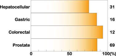
Fig.2
Increased
expression of midkine in human carcinomas. Cited from Muramatsu, T., J. Biochem.
132, 359-371 (2002)
Furthermore, midkine expression is strongly increased in all cases of Wilmsユ
tumor, in which loss of function of the tumor suppressor gene WT1 is frequently
observed, and also in all cases of malignant nerve sheath tumor (MNST) caused by
loss of the NF1 tumor suppressor gene. In neuroblastoma, urinary bladder
carcinoma and gliobastoma, patients with tumors expressing a high level of
midkine exhibit a worse prognosis than patients with tumors having a low level
of midkine.
The serum midkine level is increased in many cancer patients, and is planned to
be used as a tumor marker. The midkine level frequently increases in the early
stages of cancer progression, and is relatively high in cases of tumors with a
poor prognosis, making the midkine level a promising tumor marker.
Midkine is thought to enhance tumor progression by promoting the survival,
growth, migration and angiogenic activity of tumor cells. Antisense oligo DNA
directed at midkine suppresses growth of tumors in nude mice, opening the way to
midkine-targeted cancer therapy. Furthermore, based on the preferential
expression of midkine in tumors, the midkine promoter can be used to selectively
express toxic genes in tumors. Animal experiments have been successful.
[Inflammatory diseases and midkine ]
Midkine plays a central role in inflammation. For example, knockout mice
deficient in the midkine gene poorly develop neointima, when the artery is
damaged by ischemic shock. Renal damage after ischemia is also less extensive in
the knockout mice than in wild-type mice. Furthermore, rheumatoid arthritis in
an experimental model and adhesions after surgery are much less severer in the
knockout mice. Midkine promotes the migration of inflammatory leukocytes, namely
macrophages and neutrophils. This migration is essential for inflammation, and a
lack of midkine is considered to lead to prevention or a change of pathological
status based on inflammation. Midkine is becoming a molecular target for the
treatment or prevention of inflammatory diseases.
[Prevention of cell death using midkine]
Midkine has anti-apoptotic activity; the effect is best illustrated using
embryonic neurons as target cells. Rentinal photoreceptor cells die after
exposure to constant light in rats. Prior injection of midkine to the retina
prevents the cell death. Temporary brain ischemia in gerbils leads to delayed
neuronal death in the hippocampus. Prior delivery of midkine to the ventricle
retards this process.
Midkine is heavily deposited in senile plaques of patients with Alzheimerユs
disease. Midkine binds to amyloid b-peptide and suppresses the cytotoxic
activity. There is a possibility that midkine is produced to counteract the
toxicity of amyloid
β-peptide.
Midkine enhances the survival of bovine embryos cultured in vitro. Furthermore,
midkine suppresses infection of HIV in target cells.
The anti-apoptotic and cell-protecting activites make midkine a promising
therapeutic. However, the proinflammatory activity and protective activity of
midkine should be carefully evaluated in each case.
[Essentials of midkine]
Protein
Midkine is a basic protein, essentialy composed of two domains held together by
disulfide linkages. Each domain contains three anti-paralel b-sheets (Fig. 3).
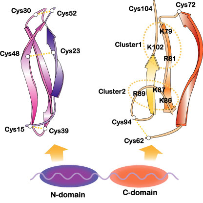
Fig. 3
The domain
organization of midkine and three dimensional structure of the domains. Two
heparin-binding sites in the C-domain are encircled. Cited from Muramatsu, T.,
J. Biochem. 132, 359-371 (2002); Wiley Encyclopedia Mol. Med. pp2086-2088 (2002)
[・2002,
John Wiley & Sons]. This material is used by permission of John Wiley & Sons,
Inc.
The more C-terminally located domain is usually responsible for midkine
activity. Midkine is dimerized through the action of transglutaminase. Some
midkine activity requires this dimerization. Pleiotrophin[also called HB-GAM (heparin-binding
growth-associeted molecule)]has 45 % sequence identity with midkine (Fig. 4).
Fig.4
Protein structure
of human midkine (MK). Amino acids conserved with pleiotrophin (PTN) are boxed.
S-S linkages are shown by lines. Arrowheads show exon boundaries. Amino acids
conserved also in Drosophila miple are shaded. Cited from Muramatsu, T., J.
Biochem., 132, 359-371 (2002)
Midkine has been found in all vertebrata examined, namely from human to
zebrafish. Zebrafish has two species of midkine. Although Drosophila lacks
midkine, miple, and miple 2 molecules with repeating units homologous to the
C-terminal half of both the midkine and pleiotrophin are present.
Gene
The human midkine gene is present in chromosome 11 band p.11.2, and is flanked
by DGKz (diacyglycerokinase z gene) and CHRM4 (muscarnic acetylcholine receptor
4 gene) (Fig.5).
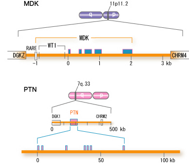
Fig.5
Structure of the
human midkine gene (MDK). For comparison, the human pleiotrophin gene (PTN) is
also shown. <,exon, RARE, retinoic acid responsive element; WT1, binding site
for WT1 protein. cited from Muramatsu, T., J. Biochem. 132, 359-371 (2002)
The symbol for the human midkine gene is MDK. The mouse midkine gene (Mdk)
is present on chromosome 2. In the upsteam of MDK, there is a retinoic
acid responsive element, and midkine gene expression is induced by retinoic
acid. Furthermore, the upstream region has a binding site for Wilmsユ
tumor suppressor WT1. When the function of WT1 is lost, suppression does not
take place, and midkine comes to be expressed. Although the pleiotrophin gene is
located in a broader region of the human genome, the fundamental structure is
similar.
Function and action mechanisms
Midkine is most strongly expressed in midgestation. Epithelial tissues involved
in epithelial mesenchymal interactions, nervous tissues during differentiation
and mesenchymal tissues undergoing remodeling are the principal sites of
expression. In the adult, midkine expression is restricted. Endothelial cells of
blood vessels and mucus epithelium of certain organs are important sites of
expression. When a tissue is injured, midkine expression is increased or newly
induced. Midkine promotes the survival and migration of various cells, and also
has many other activities (
Table 1). Using a blood vessel model, in which endothelial cells are layered
on gels with smooth muscle cells, the complex mode of midkine action during
epithelial mesenchymal interactions has been clarified (Fig. 6).
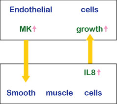
Fig.6
The action mechanism of midkine in epithelial mesenchymal interactions.
Among midkine receptors, receptor-type protein tyrosine phophafase z (PTP z) has
been studied extensively. Midkine binds to the chondroitin sulfate portion with
high affinity and to the protein portion with low affinity. In addition, low
density lipoprotein receptor-related protein (LRP) and anaplastic leukemia
kinase (ALK) have also been identified as receptors. Syndecans, a family of
transmembrane heparan sulfate proteoglycans, can also participate in midkine
signaling. The midkine receptor is considered to be a molecular complex
containing these proteins. Very recently, integrins have been found as
components of the receptor. The downstream signaling system contains PI3 kinase
followed by ERK (Fig.7).
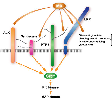
Fig.7
The signal
receptor complex of midkine (MK) and the downstream signaling system. Cited from
Muramatsu, T., J. Biochem., 132, 359-371 (2002)
Midkine binds to the oversulfated portion of heparan sulfate and chondroitin
sulfate. The structure is shown in Fig. 8
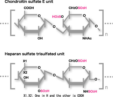
Fig.8
Carbohydrate
structure required for strong binding to midkine. The Trisulfated structure in
heparan sulfate and chondroitin sulfate E structure are shown.
Midkine is also incorporated into the cell and translocated to the nuclers. The
survival promoting activity requires the nuclear translocation. LRP is involved
in the uptake and nuclelin or laminin-binding protein precursor participates in
the nuclear translocation.
[Further information]
More detailed information is available through reading review articles or
visiting other home pages. The following are recommended.
1. Muramatsu, T. (2002) Midkine and pleiotrophin: two related proteins involved
in development, survival, inflammation and tumorigenesis. J. Biochem 132,
359-371.
[http://jb.oxfordjournals.org/cgi/reprint/132/3/359]
2. Muramatsu, T. (2002) Midkine in Wiley Encyclopedia of Molecular Medicine
pp2086-2088. John Wiley & Sons., Inc. New York, USA
3. Muramatsu, T. Chondroitin sulfate E in signaling of the growth factor
midkine. [http://www.glycoforum.gr.jp/science/glycogenes/09/09E.html]
4. Kurtz, A., Schulte, A. M., and Wellstein, A. (1995) Pleiotrophin and midkine
in normal development and tumor biology. Crit. Rev. Oncol. 6, 151-177
5. Locus link (http://www.ncbi.nlm.nih.gov/LocusLink/LocRpt.cgi?l=4192)
6. Kadomatsu, K., and Muramatsu, T. (2004) Midkine and pleiotrophin in neural
development and cancer. Cancer Lett. 204, 127-143.
7. Muramatsu T, Muramatsu H, Kaneda N, Sugahara K. (2003) Recognition of
glycosaminoglycans by midkine. Methods Enzymol. 363, 365-376.
It is also possible to read original articles listed in
Original articles as references. By
searching Pub Med [http://www.ncbi.nlm.nih.gov/PubMed/]
using midkine as a key word, more articles become available. The summary of an
article listed in
Original articles as references can be
accessed using the PMID number written at the end of each reference.
|

